
Aliviar Mezquita Arne abdominal anatomy diagram curva Nuevo significado
Female Anatomy Diagrams of the inside and outside of female body parts By Brandi Jones, MSN-ED RN-BC Updated on April 26, 2023 Medically reviewed by Lauren Schlanger, MD Fact checked by Sarah Scott Table of Contents View All Diagram External Internal Breast Anatomy Functions

Female Abdominal Anatomy TrialExhibits Inc.
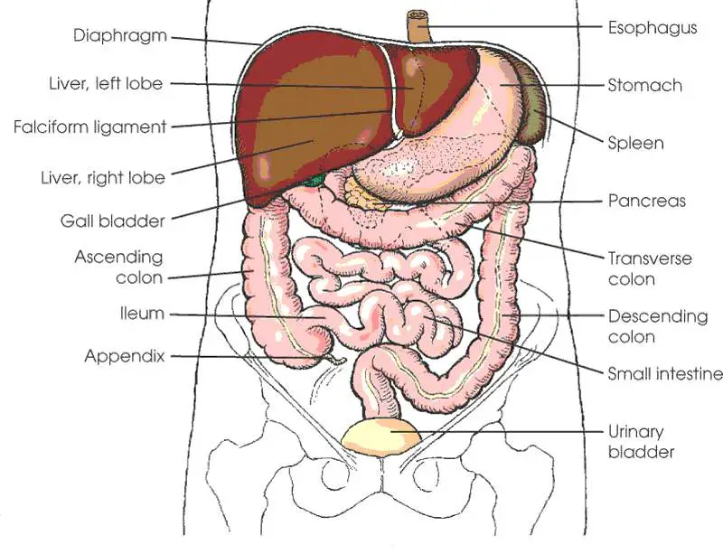
The diaphragm marks the top of the abdomen and the horizontal line at the level of the top of the pelvis marks the bottom. Connective tissue called the mesentery holds the abdominal organs together. Several large blood vessels travel through the abdomen.
Anatomy Of The Female Abdomen And Pelvis, Cut away View Healthiack
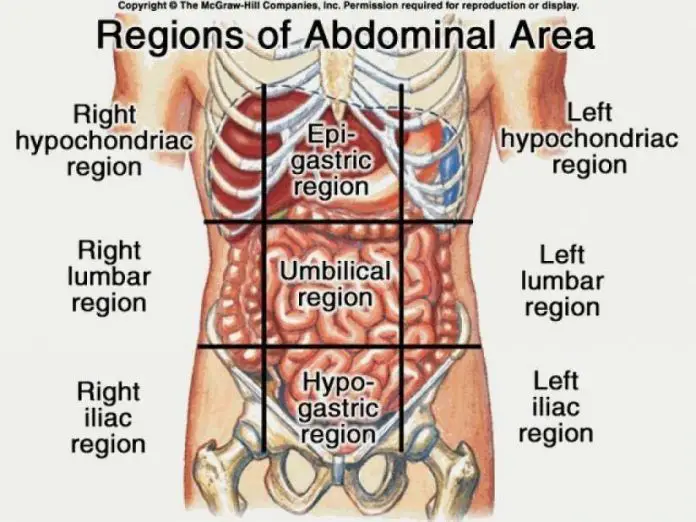
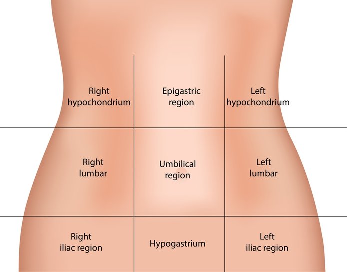
1/9 Synonyms: Abdominal region, Regio abdominis , show more. The abdominal wall surrounds the abdominal cavity, providing it with flexible coverage and protecting the internal organs from damage.

Abdominal Anatomy Pictures Female Abdominal anatomy female right side
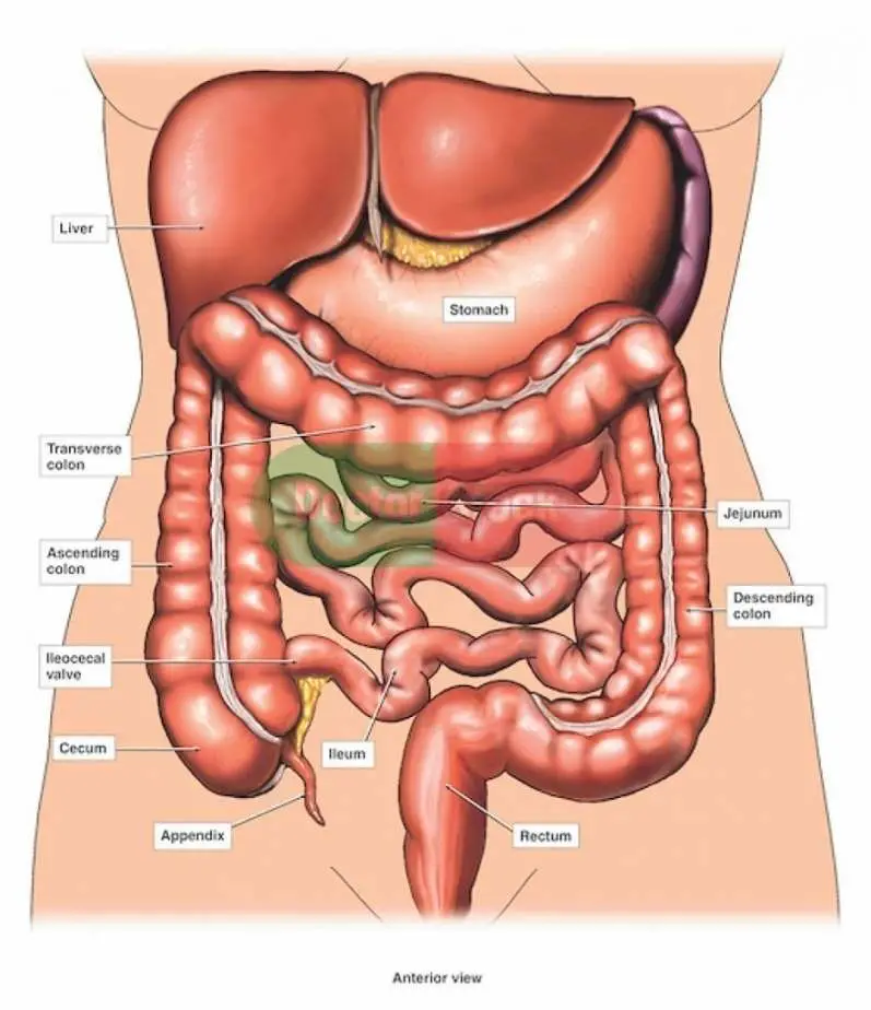
1. Anterior view: anatomy of female abdomen and pelvis: skin 2. Anterior view: anatomy of female abdomen and pelvis: muscles of anterior abdomen wall 3. Anterior view: anatomy of female abdomen and pelvis: stomach and omentum 4. Anterior view: anatomy of female abdomen and pelvis: small bowel and colon 5.

Anatomy of a Female Abdomen TrialExhibits Inc.
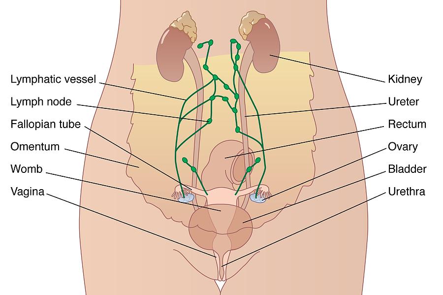
Two female reproductive organs located in the pelvis. Fallopian tubes. Carry eggs from the ovaries to the uterus. Cervix. The lower, narrow part of the uterus (womb) located between the bladder and the rectum. It forms a canal that opens into the vagina, which leads to the outside of the body. Vagina.
Anatomy Of The Female Abdomen And Pelvis, Cut away View
Anatomy atlas of the female pelvis: 101 labeled illustrations of the female genital system (ovaries, uterine tubes, uterus, vagina, vulva, clitoris) and pelvic cavity (bladder, rectum, pelvic diaphragm, perineum with innervation and blood supply). Thorax, coeur, abdomen et pelvis. Torsten B. Möller - Emil Reif. Paru le : 06/2014.

Abdominal Anatomy Male / Berlina Luwirisa Berlinaluwirisa Profile
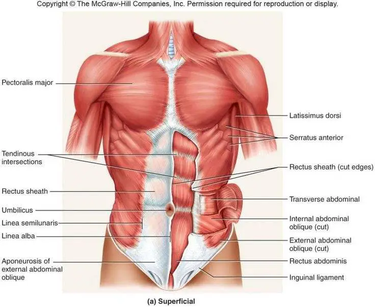
Anatomy Where are your abdominal muscles located? There are five main muscles in your abdomen. Two are vertical (up and down) muscles located toward the middle of your body. Three are flat muscles stacked on top of each other, situated toward the sides of the trunk. The two vertical muscles are:
Anatomy Of The Female Abdomen And Pelvis, Cut away View Healthiack
Abdominal Organs Anatomy, Diagram & Function | Body Maps Human body Digestive System Bones and Organs Bones and Organs At the height of the cavity is the liver, the body's largest organ. It.
Female Abdominal Anatomy, Artwork Photograph by Peter Gardiner
Human body Abdomen Abdomen The muscles of the abdomen protect vital organs underneath and provide structure for the spine. These muscles help the body bend at the waist. The major muscles of.
Abdominal Anatomy / Stomach Anatomy Photograph by Asklepios Medical
Vulva Female reproductive organs are very different to those of males. The vulva refers to the external parts of a female's genitals. It consists of several parts, including the labia majora,.
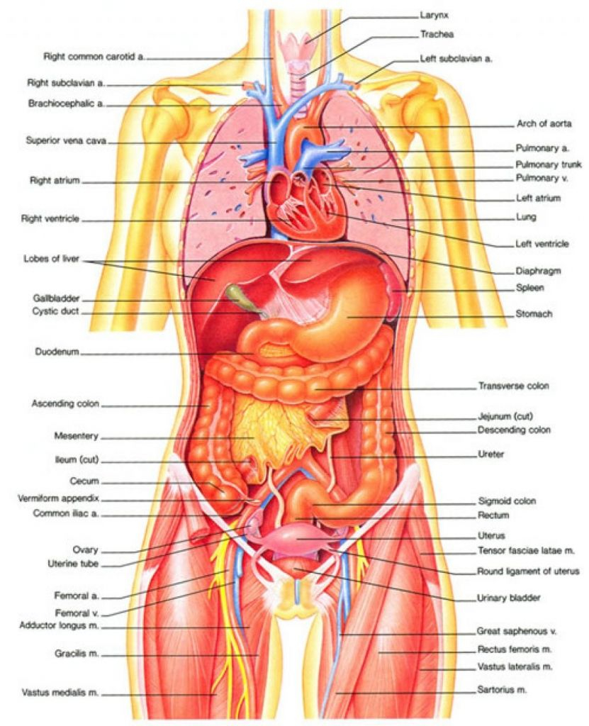
Diagram Of Abdominal Organs exatin.info
ISSN 2534-5079. This e-Anatomy illustrates the gross anatomy of the digestive system. We focused especially on the diagrams of the abdominal digestive system (oesophagus is described on the modules about the thorax and oral cavity/pharynx on the ENT modules). 84 anatomical diagrams and histological images with over 300 labeled anatomical parts.

Human Anatomy Abdomen Anatomy Pinterest
The rectus abdominis is the large muscle in the mid-section of the abdomen. It enables the tilt of the pelvis and the curvature of the lower spine. Next to it on both sides of the body is the.
Anatomy Of The Female Abdomen And Pelvis, Cut away View Healthiack
The abdomen is the part of the body that contains all of the structures between the thorax (chest) and the pelvis, and is separated from the thorax via the diaphragm. The region occupied by the abdomen is called the abdominal cavity, and is enclosed by the abdominal muscles at front and to the sides, and by part of the vertebral column at the back.
Anatomy Of The Female Abdomen And Pelvis, Cut away View Healthiack
Anatomy of the Female Abdomen and Pelvis ID: exh6130a Cite this Item Add to Collection This medical illustration depicts a mid-sagittal view of the normal anatomy of the female abdomen and pelvis. Labeled structures include the large bowel (colon or large intestine), umbilicus, small intestine, ovary, fallopian tube, uterus and bladder. Variations
Drawing Abdomen Showing Abdominal Organs Female Stock Illustration
Human body Digestive System Stomach Stomach Stomach The stomach is on the upper-left area of the abdomen below the liver and next to the spleen. It stores and breaks down the foods and.
Abdominal Anatomy Pictures Female Female Lower Abdominal Organs
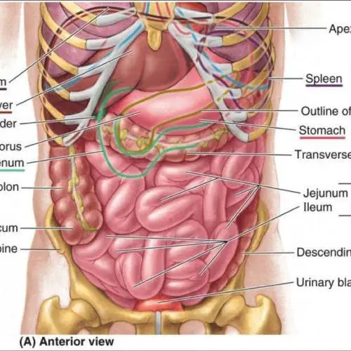
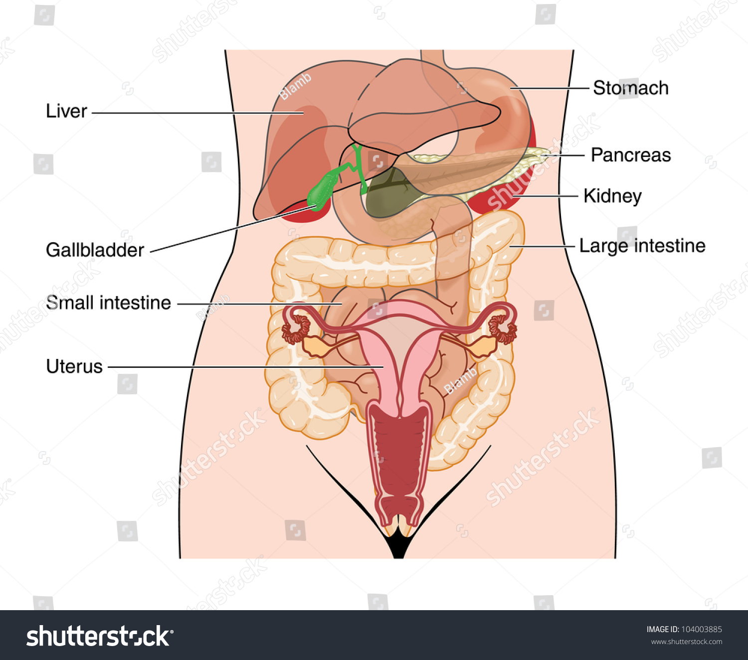
This medical exhibit diagram illustrates the anatomy of the female abdomen and pelvis from an anterior (front) cut-away view, showing elements of the digestive system. The liver, stomach, and abdominal contents are clearly identified and labeled, including the cecum, ascending colon, transverse colon, descending colon, and small intestine. The image also shows the pelvis, uterus, and urinary.